
From time to time on the Neurosurgery Blog, you will see us cross-posting or linking to items from other places when we believe they hit the mark on an issue. We wanted to bring attention to a recent publication in Neurosurgery, an official journal of the Congress of Neurological Surgeons, which provides multimedia, prompt publication of scientific articles on clinical or experimental surgery topics important for the brain, spine, and peripheral nerves, reviews, and other information of interest to readers across the world. The article, “An Endovascular Approach for Idiopathic Intracranial Hypertension: Assessing Morphometric Candidacy After Venous Sinus Stenting” is published as part of Neurosurgery’s High-Impact Manuscript Service (HIMS).
Idiopathic intracranial hypertension (IIH) is characterized by elevated intracranial pressure in the absence of a mass lesion or obstructive hydrocephalus. It predominantly affects women of childbearing age and typically presents with headache, pulsatile tinnitus, and, in severe cases, visual morbidity from papilledema. When medical therapy and weight loss fail, conventional cerebrospinal fluid (CSF) diversion remains imperfect: ventriculoperitoneal and lumboperitoneal shunts carry nontrivial complication and revision rates. Endovascular venous sinus stenting offers a less invasive option for selected patients with venous outflow obstruction, although it generally requires antiplatelet therapy and can be limited by symptom recurrence.
The CereVasc eShunt® Implant is an investigational endovascular CSF shunt designed to address some of these constraints by replicating the function of arachnoid granulations. Delivered via a venous approach through the groin, it accesses the cerebellopontine angle cistern from the inferior petrosal sinus. It drains CSF across the dura into the jugular venous system without a cranial burr hole, invasive catheters, or peritoneal tubing. Early experience in normal pressure hydrocephalus has been encouraging; although whether IIH anatomy can routinely permit analogous deployment has been less clear.
In a morphometric study published in Neurosurgery, investigators at Tufts Medical Center evaluated IPS and cerebellopontine angle cistern dimensions in 84 adults with IIH against device- and trial-informed eligibility thresholds. Under stringent, moderate, and permissive cistern depth criteria, eShunt deployment was anatomically feasible in 43%, 69%, and 82% of patients, respectively. The authors conclude that a substantial fraction of adult IIH patients meet morphometric criteria for deployment based on cistern and IPS anatomy alone, and they position endovascular CSF shunting as a potential complement—or alternative—to traditional CSF diversion and venous sinus stenting.
The eShunt® Implant System remains investigational. The anatomic feasibility data in this study does not establish clinical efficacy in IIH, but instead supports further exploratory clinical study in carefully selected patients.
To read the full Neurosurgery article, click here.
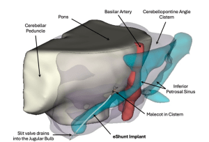
Figure 1A. Three-dimensional rendering of the CereVasc eShunt Implant in the inferior petrosal sinus, with surrounding structures at the cerebellopontine angle cistern. Reproduced from Vajipayajula DN, Yu A, Koo AB, et al. An Endovascular Approach for Idiopathic Intracranial Hypertension: Assessing Morphometric Candidacy After Venous Sinus Stenting. Neurosurgery. 2026. DOI: 10.1227/neu.0000000000004154. © Congress of Neurological Surgeons 2026. All rights reserved.


